
AI Analyzes Years of Mammograms to Better Predict Future Breast Cancer Risk
Guest Contributor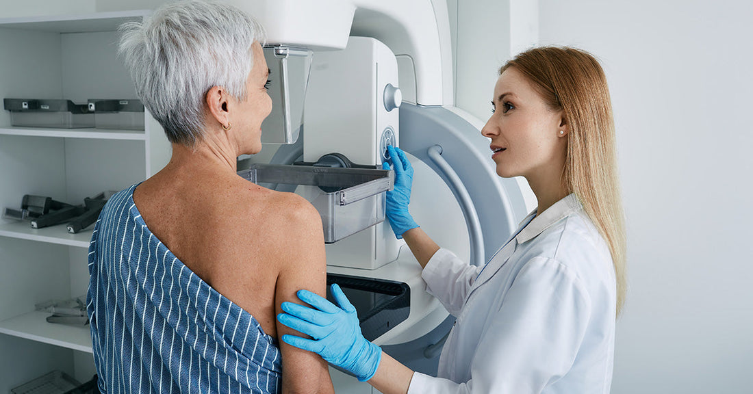
Artificial intelligence is quietly reshaping how clinicians think about breast cancer screening, and a new study from NYU Langone Health suggests that AI-assisted imaging could help personalize screening in ways that were not possible before. By using an AI model that looks at a woman’s 3D mammograms over multiple years, researchers found they could better predict five-year breast cancer risk than with standard tools that rely on a single image or non-imaging risk factors. For anyone following advances in AI and breast cancer screening, this is an important development that highlights how technology might improve early detection.
The deep-learning model at the center of the study is called NYU-DRP. It was trained to analyze longitudinal digital breast tomosynthesis, which means it evaluates a series of 3D mammograms taken annually over time. Instead of focusing on a single snapshot, the model learns from changes in breast tissue across multiple screenings. The researchers report that this longitudinal approach captures subtle patterns that traditional methods may miss, especially those based on a single 3D mammogram or on older 2D imaging methods.

In findings published online in the American Journal of Roentgenology on August 12, the team showed that NYU-DRP correctly ranked women at higher risk of developing breast cancer within five years 72 percent of the time. For comparison, a model that used only the most recent single 3D mammogram reached 70 percent, and an AI model that worked with 2D mammograms reached 68 percent. Even a few percentage points can represent many women when applied at population scale, and the improvement becomes especially meaningful when the goal is to find cancers earlier and tailor follow-up more intelligently.
The researchers built NYU-DRP using an extensive dataset: 313,531 yearly 3D mammograms from 161,165 women who did not have breast cancer at the time of imaging. All scans were performed at NYU Langone hospitals between 2016 and 2020. This large real-world dataset allowed the model to learn from a wide range of breast types, ages, and imaging patterns, while still reflecting a contemporary screening environment where digital breast tomosynthesis is increasingly common.
Study lead investigator Yanqi Xu, PhD, a postdoctoral research fellow in the Department of Radiology at NYU Grossman School of Medicine and Perlmutter Cancer Center, noted that existing 3D mammograms contain valuable information about how breast tissue evolves across time. I found this detail striking because it reframes ordinary yearly screenings as a type of time-lapse record of breast health rather than a set of disconnected exams. According to Xu, models like NYU-DRP can tap into this longitudinal information to deliver more reliable estimates of a woman’s future breast cancer risk.
The team did not stop at comparing NYU-DRP to other imaging-based AI systems. They also evaluated the model against the Tyrer-Cuzick risk assessment, a widely used tool that estimates a woman’s lifetime risk of breast cancer based on personal and family medical history, genetic mutations, breast density, and biopsy results. When the researchers focused on five-year risk, NYU-DRP again came out ahead. It correctly predicted who was at higher risk 67 percent of the time, while Tyrer-Cuzick reached 56 percent.
To make this comparison, researchers examined data from 432 women. Half of them developed breast cancer within five years, and each of these women was carefully matched to women of similar age and background who did not develop the disease in that same period. This matched design helped clarify how well each tool distinguished higher-risk individuals from those at lower risk. Importantly, less than 3 percent of women in the overall study population developed breast cancer during the trial period, which reflects the reality that most screened women will not receive a cancer diagnosis yet still undergo routine imaging and, in some cases, additional tests.
One notable finding challenges a long-held assumption in breast screening. While dense breast tissue is known to be associated with a higher risk of breast cancer, the NYU-DRP model suggested that breast density on its own does not fully capture a woman’s true risk. For women with extremely dense breasts, the model classified 37.6 percent as having average risk, and the actual cancer rate for this group after five years was 0.7 percent. On the other hand, among women with less dense, fatty breasts, NYU-DRP labeled 15.5 percent as high risk, and the actual cancer rate in this subgroup was 2.5 percent after five years.
Senior investigator Yiqiu “Artie” Shen, PhD, an assistant professor in the Department of Radiology, summarized this by saying that repeated 3D mammograms hold risk information that is not fully reflected by breast density or a single screening exam. I found it especially compelling that the model uncovered elevated risk in some women whose breasts might otherwise be considered less concerning based on density alone. This suggests that more nuanced patterns in tissue structure and change over time could matter in ways clinicians are only beginning to quantify.
The potential impact on screening strategy is clear. Study co-investigator Laura Heacock, MD, an associate professor in the Department of Radiology, highlighted that if future testing in broader populations confirms these results, AI-assisted 3D mammograms like NYU-DRP could help physicians better align screening with actual risk. Women at higher risk might benefit from more frequent imaging or supplemental tests, while those at lower risk could avoid extra procedures that add stress, cost, and exposure without clear benefit. This kind of more personalized breast cancer screening has been a longstanding goal in oncology and radiology.

The research also outlined practical next steps. Shen and colleagues plan to use the longitudinal digital breast tomosynthesis program to monitor women’s breast health prospectively and to observe which individuals eventually develop cancer. They also intend to test NYU-DRP with data from other academic health centers and from different 3D mammogram manufacturers. All the exams used in the current study were acquired with equipment from a single manufacturer, Hologic Inc. of Marlborough, Massachusetts, so external testing will be key to understanding how well the model generalizes beyond the original setting.
These developments arrive against the backdrop of significant progress in breast cancer care. In the United States, an estimated 1 in 8 women will receive a diagnosis of breast cancer over their lifetime, with approximately 382,640 women expected to be diagnosed in 2026. Early detection matters greatly. The five-year relative survival rate exceeds 99 percent when breast cancer is found in its earliest stages. Advances in both detection and treatment have helped drive survival improvements, and there are now more than 4 million breast cancer survivors in the country.
Current expert guidance recommends annual mammography screening starting at age 40, and more than 43 million mammograms were performed in 2025 alone. Integrating AI-assisted imaging tools into this already widespread practice could offer a powerful way to refine risk prediction without asking women to undergo entirely new types of procedures. Instead, the same yearly mammograms could yield richer information about future breast cancer risk, particularly when captured as 3D images and analyzed over multiple years.
The study received funding from the National Science Foundation, the National Institutes of Health, the Milstein Pilot Project Fund, the Shifrin-Myers Breast Cancer Discovery Fund, and the Manhasset Women’s Coalition Against Breast Cancer. Alongside Xu, Shen, and Heacock, the research team included Jungkyu Park, PhD, Felicia Pasadyn, MA, Qi Lei, PhD, Alana A. Lewin, MD, Krzysztof J. Geras, PhD, Linda Moy, MD, and Freya R. Schnabel, MD. Their work contributes to a growing body of evidence that AI can augment human expertise in radiology. As the technology is validated in more diverse populations and clinical environments, tools like NYU-DRP may become an integral part of how clinicians estimate risk and guide screening over a woman’s lifetime.